Silvertips vs. other Hollowpoints

I don't know about silvertips but a round my partner got me to like and now carry is Winchester SXT's. I work at night and was really impressed with the low amount of muzzle flash as well as the accuracy out of my Glock 22.
 
Odd Job,

The X-Ray pictures you linked to show how important it is to properly take contrast and dynamic range into account when performing any kind of imaging operation. Contrast and dynamic range affect visibility in any kind of imaging, be it X-ray ,visible light or thermal imaging.

Here is the X-ray you linked to now clearly showing the aluminum casing (#7). I used the auto-equalization function in the free-ware photo editing software that came on this computer to adjust your picture. It could be made much nicer to view, but I just let the photo software do it automatically rather than dink around with it manually.
attachment.php


Just a couple of minutes with an internet search engine will show that aluminum is indeed faintly radio-opaque. In fact, aluminum step wedges are commonly used to calibrate x-rays.

What's more, even soft tissues can be imaged using X-rays and the proper techniques.
 

Attachments

  • Equalized MPC  X-ray.jpg
    Equalized MPC X-ray.jpg
    221.5 KB · Views: 575
@ JohnKSa

There are a few flaws with your manipulation:

1) The black area on the original radiograph represents film that was exposed by an unattenuated X-ray beam. In other words there was nothing being traversed by the X-ray beam. For arguments sake, let us consider only the black areas where no cartridge components were present on the cassette. Your manipulation has raised black total density to a mixed black and white mottle. This is a false representation of the latent image and is not diagnostic. It is a manufactured density.

2) Your manipulation would fail if there was any overlying/surrounding anatomy. The radiograph I provided was the simplest and clearest example of how you cannot see aluminium cartridge/projectile components on a radiograph. The items were resting directly on the cassette with no composite shadowing from overlying anatomy. Radiographs for medical diagnosis don't have that base black (unattenuated areas where you produced mottle) as the general background. The background or area of interest is anatomy that has been X-rayed and attenuated the beam to some extent so that you have a density that is more grey than black. If you try that manipulation on a similar radiograph with an abdominal phantom overlying it, you'll just get a white-out. You won't see anything. In fact you'll lose the densities that we can already see on the radiograph before you manipulate it.

3) That kind of manipulation is not possible on film-based systems, which still make up the majority of X-ray imaging systems in the world today. Even on digital systems in use today, you are limited by whatever software is available on your DICOM display device. You are limited to window width and center adjustments and you cannot perform any sort of filtering or multi-step selected pixel value adjustments. The reason for this is medico-legal. It is not permitted to make a manipulation of a radiograph so that the densities of the items being X-rayed are falsely represented. This is to prevent people from 'manufacturing' or 'hiding' features that may have a bearing on the patient's diagnosis. To this end, you'll find that even the basic DICOM adjustments that are done on today's high-end systems are not one-way adjustments. There is always a reset facility so that the image can be viewed in its original state. If this was not the case a radiologist could avoid a law-suit to do with missed micro-calcs on a mammogram simply by manipulating the image so that these are no longer visible. Also don't forget that the image was already manipulated before you got it: I scanned that film here on a 4800dpi high-end scanner. It may have picked up densities that would otherwise not have been visible under other normal hospital digitisation techniques (which are typically done at 1200dpi max, and do not have any hardware filtering applied).

4) Your manipulation has failed to show the Silvertip jacket. No part of it can be seen even with your aggressive manipulation. The same can be said of the aluminium cassette surface upon which the items were X-rayed. The rear end of the cartridge case can be seen and so I must concede that the cartridge case has a latent image of itself recorded on the film. This may be more to do with other metallic components combined with the aluminium of that cartridge case being faintly radio-opaque (either by alloy or by impurity). However even so, the case is not visible on the original radiograph and I contend that it would not be possible to see it if there was overlying anatomy because your manipulation will make the overlying anatomical densities completely obliterate the cartridge case density.


Technically, everything except a vaccuum will attenuate an X-ray beam. That means that technically there is no such thing as a radiolucent structure. Sufficient quantities of a material and/or manipulation of the latent image will reveal anything you care to show (on its own) but that is outside the realms of medical imaging today. The fact remains that on X-rays for medical diagnosis you are not going to see any aluminium projectile components. There is no getting away from it. Your manipulation is impressive, but it is not available/permitted in diagnostic radiography today. More importantly though (in this case with the argument about this aluminium), it would certainly fail if there was any anatomy surrounding the area of interest.

I suspect the only way I could convince you of this is to X-ray various phantoms/blocks of meat with aluminium components embedded therein and send you the original radiographs (the original films).
 
Last edited:
Odd Job,

Yes, once an X-Ray negative has been exposed, it's not possible to make the sort of adjustment I did if you're dealing exclusively with the film. And, even with the sort of adjustment possible with computer manipulation, an improperly exposed and/or developed negative may still not show some thing that CAN be shown if the exposure and development process are managed correctly. (Like the SilverTip Jacket in this case. Although, I think if you do an actual overlay of the visible and X-Ray images, I think you will find that it is nearly all visible.)

Whoever took that X-Ray and developed it did not do so with an eye to detecting aluminum and therefore the aluminum showed up VERY poorly in some cases and not at all in other cases.

I have shown that X-Rays CAN show aluminum, as I stated in my eariler post, and what's more, I did it with your picture. Furthermore, as a pointed out earlier, it only takes a little work with an internet search engine to show that aluminim is faintly radio-opaque and that it is even used to calibrate X-rays--something that would not be possible if it were invisible.
I suspect the only way I could convince you of this...
There is no way you will convince me. In the first place, I just posted an X-Ray with aluminum visible on it. You may not like how I did it, but clearly your assertion that aluminum is invisible on X-Rays is incorrect based solely on that picture. If that weren't enough, it only takes a little bit of internet search to confirm that aluminum is indeed visible on X-Rays. I can post a bunch of links if you want, but given that you're balking at the picture, it seems a waste of my time.
Technically, everything except a vaccuum will attenuate an X-ray beam.
Not just technically, that is the absolute truth. That is why it is POSSIBLE to not only see light metals like aluminum, but also to image soft body tissues using X-Rays and the proper techniques.
Your manipulation has raised black total density to a mixed black and white mottle.
The film was not properly exposed and developed to detect aluminum. It was exposed and developed to show details in the VERY dense lead objects and therefore the aluminum was washed out. My manipulation showed that the aluminum is visible, but because of the way the X-ray was initially made and developed, the results were not ideal.
Your manipulation would fail if there was any overlying/surrounding anatomy.
It's likely that the manipulation would yield far better results if there were only overlying/surrounding anatomy. The problem with the X-Ray as it stands is that the VERY dense lead is monopolizing the dynamic range and washing out the aluminum. With only much less dense soft tissue as a background, the aluminum would be far more easily visible.
 
@ JohnKSa

Yes, once an X-Ray negative has been exposed, it's not possible to make the sort of adjustment I did if you're dealing exclusively with the film. And, even with the sort of adjustment possible with computer manipulation, an improperly exposed and/or developed negative may still not show some thing that CAN be shown if the exposure and development process are managed correctly. (Like the SilverTip Jacket in this case. Although, I think if you do an actual overlay of the visible and X-Ray images, I think you will find that it is nearly all visible.)

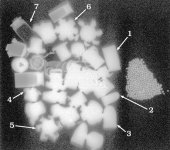
It is not. That grey halo around the bullet is not the jacket it is an anomaly associated with the lead core outline and the filter that has been applied to the image. How do I know? Because the same halo is seen associated with bullet No 3 which is an all-lead bullet. If you applied the above observation to that bullet too, you would falsely arrive at the conclusion that the bullet was jacketed. All of the high density items in your manipulated image have that grey halo, even brass cartridge bases. In fact all of them have also lost radiographic density from white to grey. This, like the mottle, is a misrepresentation of the densities on the radiograph. There are no exposure factors that can create that false representation. It is entirely due to your manipulation.

Whoever took that X-Ray and developed it did not do so with an eye to detecting aluminum and therefore the aluminum showed up VERY poorly in some cases and not at all in other cases.

I took the radiograph, and actually the converse is true. I took it specifically to image jacketing. I used the lowest radiographic exposure that we currently used in that department at the time, which is the exposure used to X-ray hands/fingers. The film/screen combination was the highest detail available in the department. It was a fine grain film with a fine grain phosphor intensifying screen. Even if we could go lower, we would not be justified to apply a non-diagnostic dose to the patient. In other words you can't justify doing a really soft X-ray exposure on a person on the off-chance that there is a piece of aluminium jacketing in a gunshot wound. It doesn't work that way. Resources wouldn't permit it even if the radiation concern was ignored. If you wanted to prospectively detect those pieces of aluminium and you had time and resources to burn, then ultrasound would be a better modality. We don't alter the X-ray exposure for forensic purposes, the exposure is always governed by clinical need.

I have shown that X-Rays CAN show aluminum, as I stated in my eariler post, and what's more, I did it with your picture. Furthermore, as a pointed out earlier, it only takes a little work with an internet search engine to show that aluminim is faintly radio-opaque and that it is even used to calibrate X-rays--something that would not be possible if it were invisible.

Yes you have shown that parts of the cartirdge case can be rendered visible under extreme and non-available/not permitted manipulation of the radiograph. If you want me to apologise to you about that, I will: here it is, I screwed up. I did not take into account the fact that the cartridge case was constructed of an aluminium alloy which would be detectable upon aggressive manipulation of my radiograph by you, JohnSKa. You showed me the cartridge case when I thought you wouldn't be able to. Does it change anythng in the world of diagnostic radiography? No, it doesn't, because in day-to-day radiography where you are X-raying anatomy there is no way you will see that. Bear in mind a cartridge case is quite big compared to a bullet jacket. Aluminium step wedges: I was the first to concede in this thread that a sizeable piece of aluminium will be seen on X-ray. I was the first to mention step wedges. If you look at a quality control film that has had a wedge exposed on it, the first step or two of aluminium are not detectable on film, and those are quite thick steps, certainly thicker than anything you would find in a projectile component. Thats why they use that step wedge: to get a range of densities from black to white. There isn't another metal you can do that with. Clearly aluminium is special in that it cannot be detected easily like other metals (and I contend that in small quantities and in the pieces of projectile fragments I have seen, it is not detectable at all with current radiography standards).

There is no way you will convince me. In the first place, I just posted an X-Ray with aluminum visible on it. You may not like how I did it, but clearly your assertion that aluminum is invisible on X-Rays is incorrect based solely on that picture.

Actually, objectively I have to agree with you. I should have been more specific about the contribution of other metals in aluminium alloys to produce a radio-opaque shadow on the film. The use of that cartridge case in this debate has been a poor choice for me. I will have to find out what its metallurgical composition is. But note that it would not have been seen in hospital viewing conditions. It is not a projectile component either. But that's a point (when argued the way you have set out above) that you must win.

If that weren't enough, it only takes a little bit of internet search to confirm that aluminum is indeed visible on X-Rays. I can post a bunch of links if you want, but given that you're balking at the picture, it seems a waste of my time.

That is not a waste of time, it just isn't relevant because it depends on which alloys of aluminium you are talking about. I think I said that in my response to Guntotin Fool above when he mentioned auto aluminium.

The film was not properly exposed and developed to detect aluminum. It was exposed and developed to show details in the VERY dense lead objects and therefore the aluminum was washed out. My manipulation showed that the aluminum is visible, but because of the way the X-ray was initially made and developed, the results were not ideal.

I used a standard hand protocol for that radiograph. In that hospital, the exposure is 50kV and 2.5mAs. It is the most unsuitable exposure to use to image lead. It is a very low exposure. That is why you can see the copper-based jackets in good detail. You cannot see any lead detail because the X-ray beam at that kVp is incapable of passing through lead. There could be a massive air bubble in all of those bullets and you wouldn't know because the X-ray beam at that kVp is stopped by the smallest thickness of lead. In fact the beam has not even been able to penetrate the bases of the brass cartridge cases. In terms of the development of the radiograph, that is automatic these days in just about every hospital you go to. Nobody dips films anymore. You don't get a processor-related change in density that is under the user's control. There is nothing wrong with the processing.

It's likely that the manipulation would yield far better results if there were only overlying/surrounding anatomy. The problem with the X-Ray as it stands is that the VERY dense lead is monopolizing the dynamic range and washing out the aluminum. With only much less dense soft tissue as a background, the aluminum would be far more easily visible.

It has nothing to do with lead monopolising anything. No density being radiographed can 'wash out' an adjacent one. In fact there is no such thing as washout in radiography. Washout applies to photography. You can't blame my radiograph for the effects of your manipulation. I have told you already, your manipulation provides a false representation of the actual densities of the items being radiographed. If this isn't the case, then how do you explain the grey halos around all the items, even the cartridge case above bullet 6 (there is no lead in that case)?

Now you've proved that some aluminium alloys can be seen in extreme circumstances. But these aren't the circumstances that apply to trauma radiography today. Even the radiograph I have posted is not indicative of the film/screen combinations employed today in the emergency room. Typical exposure values for chests and abdomens start at 70kV with 4mAs and can run to 90kV with mAs values up to 40, depending on the kV selected. At those exposures you can even miss copper-based jacketing, but you can still see tissue outlines. You can see the effects of kV increment on step wedges. At those kVs you might find that the first 4 or 5 steps of the wedge cannot be seen.
I am unmoved. Based on my knowledge of X-ray exposure factors and the appearance of projectile components in gunshot wounds I adhere to my previous statement that aluminium projectile components will not be visible on X-rays done in the hospital setting. I am still willing to prove this to anyone who doubts it. If you would like to send me your aluminium fragments I will insert those into the appropriate media and X-ray them with a range of exposures. You can have the original radiographs and tell me which ones have visible aluminium.
 
+tshadow6. I roll my own for the range or for fun on my friend's farm, .38 and .357, but when it comes to rounds for SD, I'll take factory-loaded Winchester Silvertips every day.
 
"Exposure Level #1:
X-rays can easily penetrate paper and cloth but can not penetrate denser materials such as rubber, glass, wood, or metals (aluminum, steel, and lead). "
http://www.coe.uga.edu/mse/science/outreach/xrays/x-rays.html
"To block stray radio waves emitted by the detector's circuitry, the FPD was encased in a thin aluminum skin, which is almost transparent to x rays but opaque to radio. "
http://www.aip.org/pt/vol-58/iss-6/p22.html

"A typical X-ray will penetrate through thin layers of aluminum,
and eventually be absorbed by the material."
http://www.madsci.org/posts/archives/2000-12/977443690.Ph.r.html

Quoting is hard since this is a pdf, but do a search on aluminum and find a very brief description on how aluminum is used to calibrate dental X-Rays by comparing it's known density on the images to other objects in the image.

http://www.sciencenews.org/pages/pdfs/data/1998/153-19/15319-15.pdf

"Calibration procedure of aluminum wedge by optical density gradient of its X-ray image "
http://www.ncbi.nlm.nih.gov/entrez/query.fcgi?cmd=Retrieve&db=PubMed&list_uids=2591813&dopt=Abstract

"Aluminum alloy wedges are used to standardize x-rays"

http://lsda.jsc.nasa.gov/scripts/hardware/hardw.cfm?hardware_id=777

"Aluminum FB (foreign bodies) can often be visualized on radiographs"

http://www.ncbi.nlm.nih.gov/entrez/...ve&db=PubMed&list_uids=16322976&dopt=Abstract

"A plain abdominal X-ray [Figure - 1] was advised that showed a linear radio-opaque shadow lying transversely in the pelvis. On persistent questioning, he gave a history of introduction of a 5.5 cm long aluminium rod"

http://www.jpgmonline.com/article.a...ume=46;issue=1;spage=41;epage=2;aulast=Bakshi


I think that these are sufficient to show that even standard procedure X-Rays can render aluminum visible.

However, I will concede that there are also many links indicating that locating aluminum on X-Rays can be difficult in some cases. I think that would be a much more accurate way of stating your premise since it should be abundantly clear that saying it's invisible is overstating the problem.
 
Back
Top